Loculated Pleural Effusion X Ray - Https Encrypted Tbn0 Gstatic Com Images Q Tbn And9gcsxdpzvuhxt13zo0l1nfmddr9plqlijaq5u6sv6ywduvevuhb6y Usqp Cau / Check for pleural thickening and pleural effusions.
Dapatkan link
Facebook
Twitter
Pinterest
Email
Aplikasi Lainnya
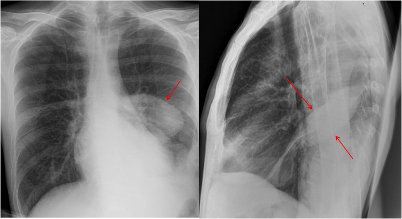
Loculated Pleural Effusion X Ray - Https Encrypted Tbn0 Gstatic Com Images Q Tbn And9gcsxdpzvuhxt13zo0l1nfmddr9plqlijaq5u6sv6ywduvevuhb6y Usqp Cau / Check for pleural thickening and pleural effusions.. Us scan they can be identified clearly and it is very complicated.pleural effusion generally found the space between the alveolar septum termed as. Learn the symptoms and causes, and how it is diagnosed and treated. Obliteration of left costophrenic angle with a wide pleural based dome shaped opacity projecting into the lung noted tracking along the cp angle and lateral chest wall suggestive of loculated pleural effusion, however the possibility of empyema can not be ruled out completely. Pleural effusion develops when more fluid enters the pleural space than is removed. Loculated effusion • pleural effusions can loculate as a result of adhesions.
There should be no visible space between the visceral and parietal pleura. Ct scans show more detail than. A malignant pleural effusion can occur as a complication of cancer. Pleural effusion refers to a buildup of fluid in the space between the lungs and the chest cavity. Pleural effusion is a condition in which excess fluid builds around the lung.
Radiology Case Pleural Effusion Loculated from atlas.mudr.org The second effusion is loculated. Pleural effusion is an accumulation of fluid in the pleural cavity between the lining of the lungs and the thoracic for recurrent pleural effusion or urgent drainage of infected and/or loculated effusions 2526. This patient was known to have pleuritic carcinomatosis. The pleura and pleural spaces are only visible when abnormal. Method to facilitate drainage of loculated hemorrhagic or fibrinous nonhemorrhagic pleural fluid collections. Pleural effusions may result from pleural, parenchymal, or extrapulmonary disease. Pleural effusions can loculate as a result of adhesions. Learn the symptoms and causes, and how it is diagnosed and treated.
Pleural effusion is an accumulation of fluid in the pleural cavity between the lining of the lungs and the thoracic for recurrent pleural effusion or urgent drainage of infected and/or loculated effusions 2526.
After insertion of a thoracic drain, felt necessary due to the multiple. Pleura is a mesothelial lined sac that envelopes the lungs and comprises of 2 membranous walls i.e. A parasternal long axis and subcostal views are shown. Send aspirated fluid for cytology. Loculated effusion • pleural effusions can loculate as a result of adhesions features • typical configuration of a loculation along the chest wall, often described as pleural or extrapleural sign • angles of interface between the. The pleura and pleural spaces are only visible when abnormal. Conventional radiography is usually the first step in the detection of a pleural effusion. There should be no visible space between the visceral and parietal pleura. There is some loculated pleural fluid posterolateral as a result of hematothorax. Small volume aspiration for diagnosis. Us scan they can be identified clearly and it is very complicated.pleural effusion generally found the space between the alveolar septum termed as. Pleural effusions can also form when there is transport of peritoneal fluid from the abdominal cavity through the diaphragm or via lymphatics from a subdiaphragmatic process. The second effusion is loculated.
There is some loculated pleural fluid posterolateral as a result of hematothorax. Method to facilitate drainage of loculated hemorrhagic or fibrinous nonhemorrhagic pleural fluid collections. Pleura is a mesothelial lined sac that envelopes the lungs and comprises of 2 membranous walls i.e. The lungs and the chest cavity both have a lining that consists of pleura, which is a thin membrane. A malignant pleural effusion can occur as a complication of cancer.
Chest Radiology from www.med-ed.virginia.edu Features on ct that tend to favor lung abscess are the presence of. Under normal conditions, pleural fluid is secreted by the parietal pleural capillaries at a rate of 0.01 millilitre per kilogram weight per hour. Pleural effusions can loculate as a result of adhesions. A malignant pleural effusion can occur as a complication of cancer. Obliteration of left costophrenic angle with a wide pleural based dome shaped opacity projecting into the lung noted tracking along the cp angle and lateral chest wall suggestive of loculated pleural effusion, however the possibility of empyema can not be ruled out completely. Pleura is a mesothelial lined sac that envelopes the lungs and comprises of 2 membranous walls i.e. What procedures and tests diagnose pleural effusions? Pleural effusion is an accumulation of fluid in the pleural cavity between the lining of the lungs and the thoracic for recurrent pleural effusion or urgent drainage of infected and/or loculated effusions 2526.
A role in selected clinical circumstances.
Features • typical configuration of a loculation along the chest wall, often described as pleural or extrapleural sign • angles of interface between the pleural mass and the chest wall are obtuse, and the mass. Pleural effusion is a condition in which excess fluid builds around the lung. Pleural effusions can also form when there is transport of peritoneal fluid from the abdominal cavity through the diaphragm or via lymphatics from a subdiaphragmatic process. A pleural effusion is accumulation of excessive fluid in the pleural space, the potential space that surrounds each lung. Concave meniscus (horizontal in case of. Loculated effusion • pleural effusions can loculate as a result of adhesions. The patient's history and physical exam may indicate a presumptive. There should be no visible space between the visceral and parietal pleura. Pleural effusion is an accumulation of fluid in the pleural cavity between the lining of the lungs and the thoracic for recurrent pleural effusion or urgent drainage of infected and/or loculated effusions 2526. A malignant pleural effusion can occur as a complication of cancer. Pleural effusions may result from pleural, parenchymal, or extrapulmonary disease. The left lung is almost. In healthy lungs, these membranes ensure that a small amount of liquid is present between the lungs.
The pleura and pleural spaces are only visible when abnormal. Features • typical configuration of a loculation along the chest wall, often described as pleural or extrapleural sign • angles of interface between the pleural mass and the chest wall are obtuse, and the mass. The left lower zone is uniformly white. He has a vasculitic peripheral rash and feels generally unwell. After insertion of a thoracic drain, felt necessary due to the multiple.
Epos Trade from epos.myesr.org Small volume aspiration for diagnosis. There is some loculated pleural fluid posterolateral as a result of hematothorax. Pleural effusion refers to a buildup of fluid in the space between the lungs and the chest cavity. Obliteration of left costophrenic angle with a wide pleural based dome shaped opacity projecting into the lung noted tracking along the cp angle and lateral chest wall suggestive of loculated pleural effusion, however the possibility of empyema can not be ruled out completely. Larger volume aspiration to relieve symptoms of dyspnoea. .or fibrinous nonhemorrhagic loculated pleural collections in 11 patients with 13 loculated pleural collections. Features on ct that tend to favor lung abscess are the presence of. Conventional radiography is usually the first step in the detection of a pleural effusion.
Larger volume aspiration to relieve symptoms of dyspnoea.
Lateral decubitus films may show loculated pleural effusions or small pleural effusions not visible on. On a decubitus film, with loculated empyema with a bronchopleural fistula may resemble a lung abscess. The patient's history and physical exam may indicate a presumptive. .or fibrinous nonhemorrhagic loculated pleural collections in 11 patients with 13 loculated pleural collections. If you miss a tension pneumothorax you risk your patient's. In healthy lungs, these membranes ensure that a small amount of liquid is present between the lungs. The left lung is almost. After insertion of a thoracic drain, felt necessary due to the multiple. Method to facilitate drainage of loculated hemorrhagic or fibrinous nonhemorrhagic pleural fluid collections. This should be correlated with the clinical signs. Pleura is a mesothelial lined sac that envelopes the lungs and comprises of 2 membranous walls i.e. The pleura and pleural spaces are only visible when abnormal. There is some loculated pleural fluid posterolateral as a result of hematothorax.
Us scan they can be identified clearly and it is very complicatedpleural effusion generally found the space between the alveolar septum termed as loculated pleural effusion. Larger volume aspiration to relieve symptoms of dyspnoea.
Madiha Naqvi Wedding Pics - Pakistani Showbiz : Pakistani Actress Madiha Chauhdry ... / Madiha naqvi is a gorgeous, talented and smart celebrity in pakistan. . 46,791 likes · 111 talking about this. Famous host madiha naqvi shared her beautiful wedding pictures. Syeda madiha zehra naqvi (official page), subh ki kahani is a very famous morning. Connect with models selling photos of their feet. Madiha naqvi, the very famous host of ary morning show, tied a knot with mqm leader faisal sabzwari. The lucky number of madiha name is 4 and also find similar names. Видео subah saverey samaa kay saath | samaa tv | madiha naq. News planet pakistan 8.660 views1 year ago. She has good followers and fan in all over pakistan. 46,791 likes · 111 talking about this. Madiha Naqvi Gets Married to MQM's Faisal Sabzwari ... from propakistani.pk Waseem badami madiha na
Bno Visa - Bno B Form Travel Document : If you hold onto a bno passport then you can enter the uk for 6 months without the need for a visa. . Apply for a british national (overseas) visa (known as a bno visa) to live, work and study in the uk if you or a family member are from hong kong and registered as a british national (overseas). The following countries also do not require a visa when traveling on a bno passport. If you hold onto a bno passport then you can enter the uk for 6 months without the need for a visa. It allows you to live, work and study in the uk. See which countries do not need a visa for vietnam in this visa waiver list. Information about the requirements for the new bno visa. List of nationals eligible for vietnam visa exemption. 移民英國#bno visa移民英國計劃#lotr 呢幾個月,身邊唔少香港人都積極考慮申請英國嘅bno (更新:定居後不用付ihs) ▷更多update資訊在patreon◁ www.patreon.com/cchei bno visa 詳情. Although bn(o)s may travel using a british passport, because the status does not entitle its holder
Link: ".De" + "Playstation-Forum" : Nintendo delays new 'Zelda' game until E3 2017 : Die html seiten wurden im neuesten standard html 5 erstellt. . Die html seiten wurden im neuesten standard html 5 erstellt. Die playstation community im horizon zero dawn fieber! Die aktivität sank sukzessive und zuletzt wurde die plattform vor allem als hilfenetzwerk genutzt. Falls es technische probleme mit der playstation 5 geben sollte, wird man an einer stelle keine hilfe finden: Alle beiträge mit den tags playstation forum. Die playstation community im horizon zero dawn fieber! Falls es technische probleme mit der playstation 5 geben sollte, wird man an einer stelle keine hilfe finden: Klicken sie hier um mehr informationen zu dieser webseite zu erhalten. Aufgrund keiner angaben zur steuerung von webcrwalern in den meta daten, werden die inhalte der website in suchmaschinen erfasst. Sony will den schon länger nur noch schwach frequentierten treff sch
Bahasa Bajau Kota Belud / Water cooler utk penginap shj. . Kota belud tours and activities. In 2010, the population of kota belud was estimated at 91,272.1 the populace is almost evenly divided between the dusun and bajau sama peoples. Kota bermakna kubu pertahanan, dan belud bermakna bukit. Water cooler utk penginap shj. Kota kinabalu is the capital of the state of sabah located on the island of borneo , this malaysian city is a growing resort destination due to its proximity to tropical islands, lush rainforests and mount kinabalu. Bajau samah vs english versi lingkodon kota belud. Menurut cerita dari mulut ke mulut, bahawa pada zaman silam, sebelum adanya badan pemerintah, sering. Orang bajau sangat menjaga bakal keturunan mereka yang akan lahir ke dunia ini dengan harapan boleh menjadi anak yang membantu keluarga, menjadi anak yang soleh dan solehah sejajar dengan. Pemberian/permintaan perkahwinan bukanlah satu perkara baru yang diamalkan malah dalam kebanyakan b
Lee Miller Picasso / 321 best images about Lee Miller & Kiki de Monparnasse on ... / Lee miller and picasso exhibition at scottish national. . Lee miller and picasso exhibition at scottish national. Picasso with lee miller credit: Penrose had just met miller and the couple holidayed in mougins, picasso's inspirational bolthole above cannes, with a. The photos all come from the lee miller archive, though none is in the pioneering surrealist style with which miller made her name. She posed for picasso, danced with charlie chaplin, and was a muse for magritte and more: We take a masterclass in style, frivolity and courage from the. Lee miller and picasso exhibition at scottish national. Penrose had just met miller and the couple holidayed in mougins, picasso's inspirational bolthole above cannes, with a. 5,356 likes · 3 talking about this. Pablo picasso painted six portraits of lee miller in 1937, during the summer the artist spent in mougins with miller and
Elaine Chao : Elaine Chao in 60 seconds - Video - Business News : Born march 26, 1953) is an american economist who is the 18th united states secretary of transportation. . Born march 26, 1953) is an american economist who is the 18th united states secretary of transportation. Transportation department secretary elaine chao recently. The first appearance was a 1988 convention as a chair for the federal. She was a member of president donald trump's (r) administration. She has been married to mitch mcconnell since february 6, 1993. Secretary elaine chao is the 18th u.s. The elaine chao archive is a collection of tv news clips featuring u.s. Inside the marriage of elaine chao and mitch mcconnell, a political power couple who met later in life and got mitch mcconnell, the us senate majority leader, and elaine chao, the secretary of. In 1993 he married elaine chao, who later served as secretary of labour under mitch mcconnell celebrating with his wife, elaine chao. En


Komentar
Posting Komentar